Description
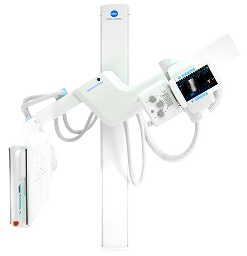
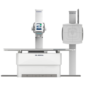
The KDR® Advanced U-Arm System employs a state-of-the-art, 17”x17” Cesium iodide (CSI) detector that maximizes efficiency and delivers image quality with excellent bone and soft-tissue visualization from a single study exposure, giving you the clarity to reach diagnoses sooner. The system maintains alignment between the X-ray tube and DR panel, regardless of panel angle or swivel arm tilt position. A durable, protective panel enclosure also helps to increase system longevity and stability.

PATIENT CENTRIC DESIGN
 KDR® Advanced U-Arm System automatically swivels into position across a 135˚ range of motion and 35″ of vertical movement, saving valuable time. The advanced detector and Ultra® acquisition software work quickly in unison to enhance the acquired image. Information is instantly retrieved, prompting the system to move to a predetermined position and source-to-image-receptor distance (SID) to save time when positioning the patient.
ERGONOMIC
Exams are simplified when the operator can confirm the patient’s information on the tube-mounted or remote console. After exposure, the image is captured and displayed for on-screen review within three seconds. Upon approval, the automated preparation begins for the next exam, minimizing time between patients.
PRECISE CONTROL
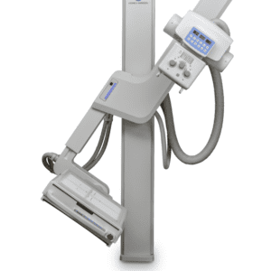
KDR® Advanced U-Arm can move from PA to Lateral positions without moving the patient and Independent SID control on the tube and the detector to support all other imaging views commonly required in radiology, including wheelchair and table work.
AUTOMATIC STITCHING
KDR® Advanced U-Arm offers optional automatic stitching with a three-knob collimator that provides precise manual control over the imaging area to reduce radiation scatter.
| X-Ray system | |
| System Type | Floor Mounted U-Arm |
| Automation | Auto positioning pre-programmed U-Arm positions |
| Generator and Tube | Multiple configurations available |
| High Speed Motorized Movement | 1. Source to Image Distance (SID) 2. Arm Elevating Movement: Detector Arm/ Tube Arm 3. Rotation of Arm & Tube 4. Rotation of Bucky |
| Source to Image | 1000~1800mm (39.5~71 in.) |
| Vertical Travel | 40~170 cm (15.7~66.9 in.) |
| Arm Rotation Angle | -30°~120° |
| Tube Rotation | ±90° |
| Detector Rotation | ±45° |
| Motion Control | Tube head, detector and wireless remote control |
| Detector and Subsystems | |
| Built-in Detector | 17”x17” amorphous silicon digital detector |
| Scintillator (fluorescent substance) | CsI (Cesium iodide) |
| Detection Quantum Efficiency (DQE) | 65% @ 0.0 cyc/mm |
| Pixel Size | 175 um |
| A/D Conversion | 16 bit (65,536 gradients) |
| Dynamic Range | 4 digits |
| Image Area Size | 425.25 x 425.25 mm (2430 x 2430 pixels) |
| Preview Display Time | under 3 seconds |
| Exposure Interval (cycle time) | 10-12 seconds (wired connection) |
| Ultra Workstation | |
| System Platform | Desktop workstation with touchscreen – duplicate screen on tube head
DICOM 3.0 Compliant features – imaging, annotation, and analysis tools |

